New paper: Overestimation of the Apparent Diffusion Coefficient in Diffusion-Weighted Imaging Due to Residual Fat Signal and Out-of-Phase Conditions
Congratulations to Maher Dhanani, Dominika Skwierawska and co-authors on the recently published article ‘Overestimation of the Apparent Diffusion Coefficient in Diffusion-Weighted Imaging Due to Residual Fat Signal and Out-of-Phase Conditions’!
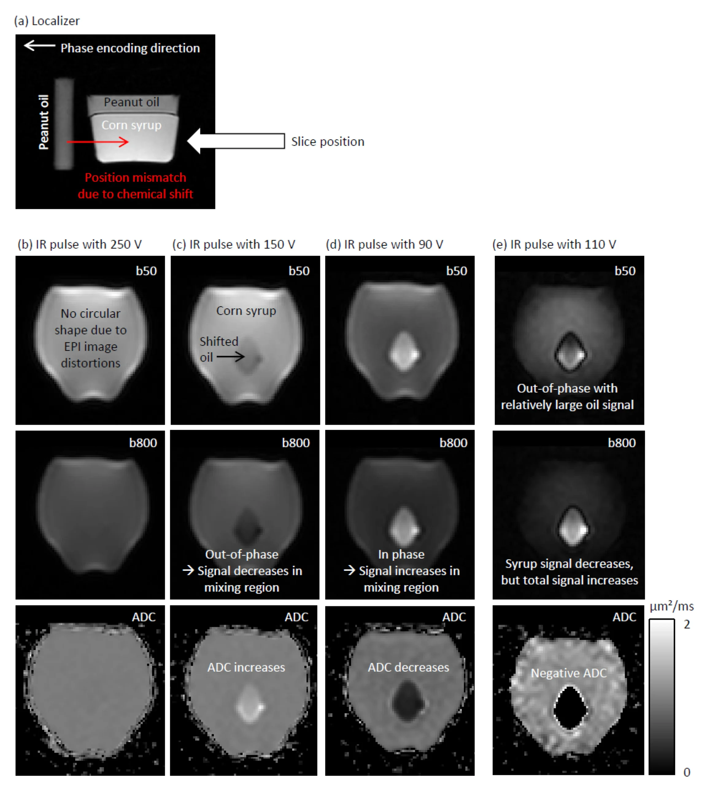
This study demonstrates that diffusion-weighted MRI can lead not only to ADC underestimation but also to ADC overestimation when residual fat and water signals are out of phase. Using fat–water phantoms and breast MRI data from healthy volunteers, they show that imperfect fat suppression can artificially increase ADC values. This effect may cause false-negative classification for lesions.
The full article can be found here: